
Comment:
I admit I love my UV, especially since moving back to New England – I have 3 apps on my phone so I know when it’s peak time to go outside. I also recognize that I have skin that tans and rarely burns, whereas other people don’t. It’s always been suspect to me that something as ubiquitous historically as sun exposure could be that bad, and data on melanoma and sun exposure does call that into question, often showing an inverse relationship. I recommend to patients to go out very early in the season and begin to build up a tan, similar to how you’d approach exercise when you’re out of shape. Like the weekend warrior exerciser, acute large sun exposure is clearly damaging. However, repetitive, low UV doses, which lead to tanning and protection is a different issue.
The concept of a “photo-induced alert state” emerging from this pre-clinical research offers a biologically optimistic perspective on our historical relationship with the sun. This study in mice suggests that this ancestral exposure paradigm isn’t simply a slow path to damage, but a mechanism of photoadaptation. The repetitive, sub-erythemal exposure seems to prime the skin’s innate immunity by triggering epidermal thickening and the intense upregulation of Antimicrobial Peptides creating an enhanced physical and chemical barrier without inducing overt inflammation.
This finding provides a clear, mechanistic basis for why “sun-hardened” skin is a real phenomenon and suggests that, within a safe threshold, the sun acts as a sophisticated immune regulator and barrier fortifier. Rather than seeing all sun exposure as a purely pathological assault, we can now appreciate that low-level, consistent UV exposure may be integral to maintaining a robust, “alert” innate immune function in the largest organ of the body. This is a critical distinction that supports the safe use of controlled phototherapy and re-contextualizes the long-term, non-inflammatory benefits of natural UV exposure.
The Wonk Debate – Audio Critique & Clinical Commentary:
Summary:
Clinical Bottom Line
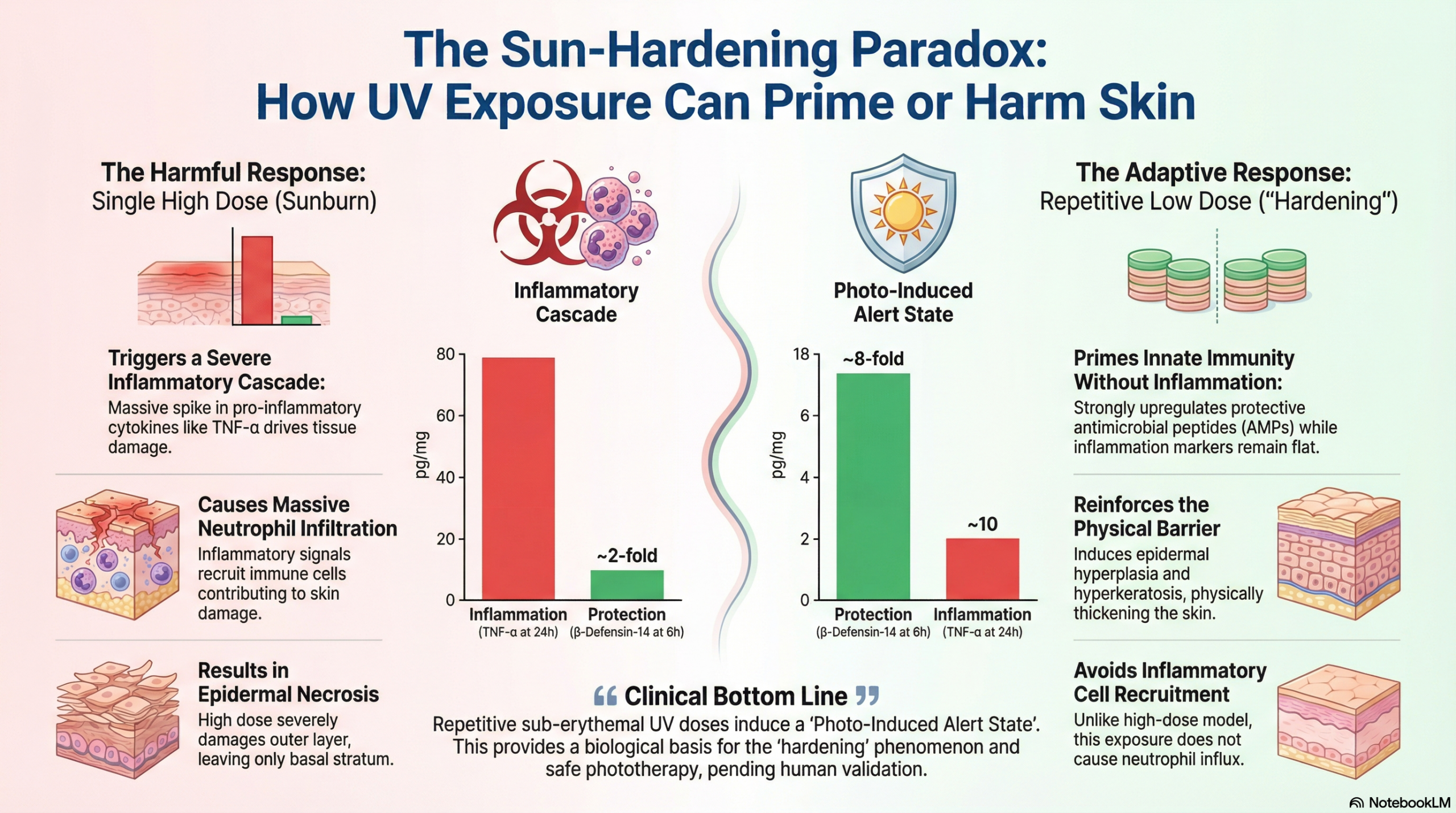
This pre-clinical study in hairless mice provides a mechanistic distinction between acute, harmful UV exposure (sunburn) and repetitive, low-dose exposure (photoadaptation). The findings suggest that while a single high dose drives severe tissue damage and a classic inflammatory cascade (neutrophil/monocyte recruitment), repetitive low doses induce a “photo-induced alert state”. This state is characterized by epidermal hyperplasia (thickening), enhanced barrier function, and the upregulation of antimicrobial peptides without overt inflammation. For the clinician, this elucidates the biological basis of skin “hardening” and suggests that low-level UV exposure primes innate immunity rather than suppressing it or causing damage.
Results in Context
Primary Outcome: Histological and Inflammatory Response
- Single High UV Dose (shUVd): This exposure (400 mJ/cm2, approx. 2 MEDs) resulted in severe epidermal damage, leaving only the basal stratum intact by 24 hours. This was accompanied by a deep inflammatory state, characterized by significant production of pro-inflammatory cytokines (TNF-\alpha, IL-6) and chemokines (CXCL-1, CXCL-2).
- Repetitive Low UV Doses (rlUVd): This exposure (4 x 20 mJ/cm2, approx. 0-1 MED/day) resulted in a hyper-proliferative state. The epidermis showed significant thickening (hyperplasia) and hyperkeratosis but no signs of inflammation or tissue necrosis.
Key Secondary & Specialized Outcomes
- Immune Cell Recruitment:
- shUVd: Triggered a massive influx of neutrophils and macrophages into the dermis, forming granulomas.
- rlUVd: Did not induce neutrophil recruitment. Instead, it maintained a stable immune environment, supporting the concept of an “alert” rather than “alarm” state.
- Antimicrobial Peptides (AMPs): The repetitive low-dose group showed a rapid and intense induction of mRNA transcription for \beta-defensin 14 (\beta-Def-14) and cathelicidin (CRAMP) in both the epidermis and dermis. The authors posit this upregulation prepares the skin to defend against potential infection during the vulnerability of UV exposure.
- Lymphatic Function:
- rlUVd: Induced VEGF-\alpha transcription (an angiogenic and lymphangiogenic factor) and led to the visualization of dilated, functional lymphatic vessels without leakage.
- shUVd: Produced less pronounced lymphatic changes and was associated with damage rather than functional expansion.
Assertive Critical Appraisal
Risk of Bias & Study Limitations
- Animal Model Translatability: The study utilizes Crl:SKH1-hrBR hairless mice. While this is a standard model for photoimmunology, murine skin differs anatomically and immunologically from human skin (e.g., thickness, hair follicle density, presence of specific immune cell subsets). Therefore, the “photo-induced alert state” must be validated in human clinical samples.
- Sample Size: The study used a relatively small sample size (n=5 per group). While statistical significance was achieved (P < 0.05), small sample sizes in animal studies can lead to overestimated effect sizes.
- Scope of Analysis: The study focused on transcriptional profiles (mRNA) and some protein quantification. While functional assays (e.g., bacterial challenge to test the efficacy of the AMPs) would have strengthened the “alert state” hypothesis, they were not performed.
Reporting Quality Assessment
- Clarity: The methodology is well-detailed, including specific primer sequences and antibody sources, allowing for reproducibility.
- Statistical Rigor: The authors appropriately used ANOVA with post-hoc tests (Student-Newman-Keuls or Dunn’s) to account for multiple comparisons across time points.
Applicability
- Clinical Relevance: These findings are highly relevant for understanding the mechanisms of phototherapy (e.g., narrowband UVB for psoriasis/eczema), where low, sub-erythemal doses are used to induce remission. The upregulation of AMPs and barrier thickening explains why conditioned skin is more resistant to infection and damage than unexposed skin.
Research Objective
The objective was to compare the impact on skin innate immunity of two contrasting UV exposures: a single harmful dose (simulating sunburn) versus repetitive low doses (simulating daily incidental exposure), specifically analyzing histology, inflammatory recruitment, and mitochondrial function.
Study Design
- Type: Pre-clinical, controlled, in vivo time-course study.
- Intervention:
- Group 1 (shUVd): Single exposure to 400 mJ/cm2 (harmful dose).
- Group 2 (rlUVd): Four consecutive daily exposures of 20 mJ/cm2 (sub-erythemal dose).
- Control: Non-irradiated, age-matched mice.
- Time Points: Analyses performed at 2, 6, 24, and 192 hours post-irradiation.
Setting and Participants
- Subjects: Male Crl:SKH1-hrBR hairless mice, 7–9 weeks old.
- Setting: Controlled laboratory environment with 12-hour light/dark cycles.
Bibliographic Data
- Title: Time-course study of different innate immune mediators produced by UV‐irradiated skin: comparative effects of short and daily versus a single harmful UV exposure
- Authors: Cela EM, Friedrich A, Paz ML, Vanzulli SI, Leoni J, González Maglio DH
- Journal: Immunology
- Year: 2015
- DOI: 10.1111/imm.12427
Fair Use & Copyright: This post provides a transformative, thesis‑driven critical appraisal intended for educational and scholarly purposes. It is not a reproduction of, nor a market substitute for, the original research article.
Support the Version of Record: To support the copyright holders and verify the underlying data—including primary survival curves, risk estimates, and other core outcomes—readers are strongly encouraged to access the original Version of Record via the link or DOI provided above.
Medical Disclaimer: This content is for informational and educational purposes only and is not a substitute for professional medical advice, diagnosis, or treatment.